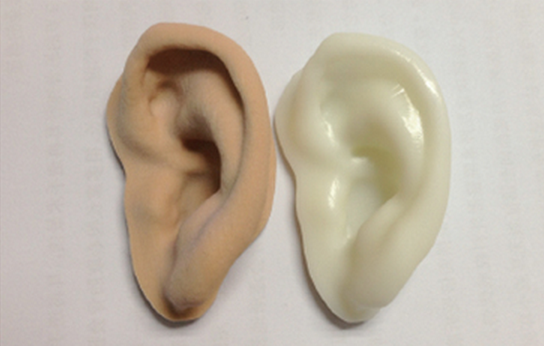
Microtia syndrome, also known as congenital microtia, is a congenital dysplasia of the auricle, often accompanied by external auditory canal atresia, middle ear malformation and maxillofacial deformity. The incidence rates vary from race to race. According to the development of auricular auricles, some or all of the ear auricles need to be reconstructed. Ear reconstruction is a difficult and complicated procedure in which the choice of ear support is the key to ear reconstruction. At present, autologous costal cartilage is considered to be the most reliable and desirable method for ear scaffolding. The use of autologous tissue for ear reconstruction is still the mainstream of ear repair. However, there are major disadvantages such as large surgical trauma, damage to the donor area, and unsatisfactory appearance. Therefore, the development of non-invasive, non-immunological rejection, fine personalized ear reconstruction surgery is an urgent clinical need. 1. Ear reconstruction surgery and ear support selection Microtia syndrome, also known as congenital microtia, is a congenital dysplasia of the auricle, often accompanied by external auditory canal atresia, middle ear malformation and maxillofacial deformity. The incidence rates vary from race to race. According to the development of auricular auricles, some or all of the ear auricles need to be reconstructed. Ear reconstruction is a difficult and complicated procedure in which the choice of ear support is the key to ear reconstruction. At present, autologous costal cartilage is considered to be the most reliable and desirable method for ear scaffolding. The use of autologous tissue for ear reconstruction is still the mainstream of ear repair. However, there are major disadvantages such as large surgical trauma, damage to the donor area, and unsatisfactory appearance. Therefore, the development of non-invasive, non-immunological rejection, fine personalized ear reconstruction surgery is an urgent clinical need. The treatment of congenital microtia is complicated, and there are various clinical surgical methods in the past, mainly including staged surgery and stage I surgery. The choice of ear stent is the key link of ear reconstruction surgery. It is the basis of auricular reconstruction. According to the choice of ear stent, it can be divided into the following three categories: one is to repair with autologous tissue and stent; the other is to use artificial material or allogeneic (species) Cartilage is used to repair the ear stent; the third is to use the deaf complex to perform ear repair. The use of deaf complex for ear repair is an ancient method. Although the surgical trauma is small and the scope of application is wide, the deafness is “non-selfâ€, the patient is not psychologically accepted, and needs to be cleaned every day. The color is hard to match with the surrounding skin, and there is even the possibility of falling off. Allogeneic ear cartilage, costal cartilage, and bovine cartilage have been used as ear supports, but they have been abandoned due to obvious disadvantages. The use of artificial materials for ear stents, such as silicone, Medpor ear stents, while avoiding surgical trauma and complications of autologous costal cartilage, has its own shortcomings. Silicone prostheses have the disadvantages of soft texture, difficult shape, incompatibility with tissues, easy rejection, fibrous envelope formation and collapse. Medpor is easy to shape and can be spliced ​​into different sizes and shapes. It has strong stereoscopic effect, no antigenicity and toxicity. A large amount of soft tissue can grow after implantation. However, the material of the stent is hard, and the stent removal rate is still high. Once the stent is exposed, it is difficult for the wound to heal itself. At present, the use of autologous tissue ear reconstruction is still the mainstream of ear repair. The ear scaffold is carved from the autologous costal cartilage. It has no immune rejection and moderate texture. The re-created skin has the same color as the surrounding skin and has a good feeling. Tanzer, Brent, Japan Fukuda, and Nagata stage auricular reconstruction surgery all use autologous costal cartilage, and achieved good clinical results. However, its shortcomings are that the surgical trauma is large, there is damage to the donor site, and it may cause thoracic deformity and severe scarring in the back of the ear. With the continuous advancement of medicine and materials science, scholars are constantly striving to find the ideal ear stent material and surgical methods. The emergence and development of tissue engineered cartilage has made it possible to solve the problem of ear reconstruction. The key issues in tissue engineering are seed cell source and carrier properties. The selection of seed cells, the establishment of a three-dimensional complex composed of cells and bioscaffold materials, and the location of growth and metabolism for seed cells are two important aspects. Cao et al. used a polyglycolic acid-polylactic acid template to shape the shape of a 3-year-old child's ear, and inoculated chondrocytes isolated from bovine articular cartilage into polyglycolic acid. The polylactic acid template was transplanted into the back of nude mice for 12 weeks in vitro, and new cartilage formation was observed by morphology and histological analysis. SH Kamil et al. (2002) used a combination of chitosan and gelatin networks with polylactic acid to prepare a cartilage scaffold material. There are also many studies in China that use different seed cells to obtain tissue-engineered cartilage. These studies offer potential applications for clinical surgery in patients with auricular defects, but more research is needed before applying tissue-engineered cartilage to clinical practice. 2. 3D printing technology 3D printing (3D Printing), also called rapid prototyping (rapid prototyping, RP), or for additive / additive manufacturing (additive manufacturing, AM), is a combination of digital, intelligent manufacturing and materials science, computer-based three-dimensional digital imaging technology And an emerging application technology for multi-level continuous printing technology. 3D printing technology was launched in the late 1980s and was originally used in manufacturing engineering and aerospace model design. With the development of 3D printing technology, some epoch-making 3D products have appeared in the fields of industrial manufacturing, culture and art, aerospace and bioengineering. 3D printing technology has received wide attention due to its high precision, short production cycle, and ability to meet individual requirements. Currently commonly used 3D printing technologies include stereolithography ( SLA ), fused deposition modeling ( FDM ), selective laser sintering (SLS) and two dimensional printing (3DP printing). )Wait. With the continuous development of 3D printing technology, this emerging scientific and technological achievements have gradually entered the medical field. The combination of 3D printing technology and medicine has become a milestone in medical history, and has been widely used in medical model manufacturing and surgical analysis planning, regeneration and repair, organ transplantation and drug development and testing. 3. Development and application of 3D bio-printing technology 3D-bioprinting is a three-dimensional computer model based on software layered discrete and numerically controlled methods to locate and assemble biomaterials or living cells, and to manufacture biomedical implants, tissues, organs and medical aids. 3D printing technology for products. 3D bioprinting can be divided into four levels: in vitro model making, permanent implantable character manufacturing, cell indirect assembly manufacturing, and direct cell assembly manufacturing. The 3D bioprinting core technology is cell assembly technology, ie cell 3D printing technology, which is a new technology for manufacturing tissue or organ precursors by locating living cells/material units. Its greatest advantage lies in the integrated manufacturing of complex shapes and internal microstructures, enabling individualized production of various organs for specific patients and specific needs. The application of 3D printing technology in orthopedic surgery can make the operation more precise and personalized, improve the success rate of complex surgery, shorten the operation time and make the operation safer. For the first time, Stoker et al. used a 3D printed model for preoperative simulation of craniofacial surgery. Levine et al. performed the surgical CT simulation of the CT data collected before surgery, and obtained information such as the osteotomy line and the target position of the bone movement, and used the position information in real time to guide the operation. With the development of materials science, research has attempted to replace the previous model materials with biological materials, and after processing by CAD software, the human implants are directly printed. Saijo et al. used a biomaterial such as tricalcium phosphate powder to prepare a personalized prosthesis. After sterilization, it can be directly implanted into the human body without engraving. The 3D printing technology is expanded from simple model manufacturing to bio-manufacturing. Kozakiewicz and others used 3D printed titanium alloy implants to repair the fracture of the sacral floor and obtained good fixation and fit. In addition, 3D bio-printing technology is also widely used in drug development. In 2012, the first 3D printed liver tissue product appeared for drug testing; in 2013, the US ORGANOVO company successfully printed a small liver tissue with normal liver function, which has the protein to transport salt, hormones and drugs to the body. The Wake Forest Institute for Regenerative Medicine will place multiple types of kidney cells cultured in living tissue in a 3D bioprinter while using biodegradable biomaterials as a scaffold. Human kidneys. The use of 3D technology to print human liver, kidney and specific cell tissue for new drug testing can not only realistically simulate the response of human tissue to drugs, but also greatly reduce the cost of research and development of new drugs. 3D cattle printing is a new type of tissue engineering technology. It can not only construct tissue engineering scaffolds with complex shapes and structures, but also realize three-dimensional precise positioning of seed cells of different densities in different scaffold materials to achieve synchronization of cells and biomaterials. Printing, and ultimately the construction of bionic tissues and organs, also known as tissue printing or organ printing, is a new breakthrough in organ transplantation and regeneration. Boland et al. The 3D printing technology was used to simultaneously print bovine vascular endothelial cells and alginate hydrogel to form a three-dimensional composite of endothelial cells and hydrogels. The active micro-vessel structure was successfully printed, which laid a foundation for printing blood vessels. Huang et al. of the University of Tokyo, Japan, used avidin-biotin to print a branched vascular system and implanted liver cancer cells thereon. In 2013, 3D printed skin and kidney research made a breakthrough in the United States. In 2014, Lee and others used 3D bio-printing technology to directly print human skin grafts, avoiding the adverse consequences of skin graft surgery. Researchers at Wake Forest University in the United States hope to print skin tissue directly on the wound site and try to build a portable printer that can be used in battlefields and disaster areas. The researchers scanned a patient's wound with a 3D bioprinter to determine the location and extent of the skin graft. Subsequently, one inkjet valve ejected thrombin; the other inkjet valve ejected cells, collagen, and fibrin. a mixture of the original components; then, a layer of human fibroblasts is printed and a layer of keratinocytes is printed. Using 3D bio-printing technology to obtain organs that are completely matched with the patient's autologous cells will bring hope to the medical community and patients. Application prospect of 4.3D bio-printing technology in ear reconstruction surgery Congenital microtia auricular reconstruction is a common organ repair operation in plastic surgery. How to use autologous tissue for ear reconstruction surgery, while avoiding surgical trauma and complications, can achieve a fine and personalized surgical effect. 3D bioprinting technology is expected to achieve this in the near future. In 2013, researchers at Cornell University in the United States used bovine ear cells to print artificial ears. When printing the ear mold, a gel that can inject collagen and living cells is borrowed, and then the printed ear mold is removed and incubated in a cell culture dish, and the cartilage can replace collagen in 3 months. Lee et al. produced artificial ears including regenerated cartilage and adipose tissue by 3D printing technology, and polycaprolactone (PCL) and a cell-filled hydrogel in a three-dimensional network structure are the main components. Hydrogels for organ 3D printing, such as sodium alginate, collagen, and block copolymers (pluronies), have low mechanical properties and instability during culture. Mannoor et al.'s research proposes the fabrication of biomimetic ears that are both complex and biologically and nanoelectronically functional through three-dimensional printing; their research proposes a new strategy for printing radios through biological cells and nanoparticles from electronic components. Frequency reception exhibits enhanced auditory perception; this bionic human ear with both fine form and function brings new hope to patients undergoing ear reconstruction surgery. 5. Outlook Through 3D bio-printing technology, biocompatible cells, scaffold materials, growth factors, signal molecules, etc. can be printed under the computer command to print physiological organs with physiological functions, which can be repaired or replaced. It has extremely far-reaching effects in the biomedical field. significance. With the research and development of 3D bio-printing technology and organ printing technology, this technology is expected to successfully print bionic tissues and organs, completely solve the limitations and problems of autologous or allogeneic transplantation, such as surgical trauma, insufficient organ source, Rejection, etc., bringing the development of application of regenerative restoration medicine and organ transplantation to a new era. Synthetic Crochet hair extensions are 100% handmade crochet fabrics, prefabricated loops and high-quality synthetic fibers.
Crochet hair of different lengths, suitable for girls, children, women. With different colors to choose from, the maintenance cost of crochet hair is relatively low, and it is an excellent style for your daily styling.
Professionally and specially weave the hair end and it is not easy to fall apart, so long-last, install it directly, save more time and money, easy to air dry naturally. Crochet Hair Extensions,Spring Twist Crochet Hair,Passion Twist Hair,Synthetic Crochet Braids Xuchang Le Yi De Import And Export Trade Co., Ltd. , https://www.synthetichairs.com
Crochet hair is made of high-quality flame-retardant low-temperature synthetic fiber. Synthetic crocheted hair is smooth, soft, durable, natural in texture, and very comfortable to wear.
How many crochet braids you need depends on whether your hair is big or small. If you need to look full, there are 8 packs for you. If you want naturalness, 6 packs are fine.